 |

Citrus Canker In-Depth

I. Economic Hosts

II. Pathogens

III. Disease

IV. Symptoms and Signs:

Citrus canker disease has been historically described as having different "forms",
including Asiatic citrus canker, false citrus canker and Mexican lime cancrosis.
However, these three "forms" are not distinctive in terms of disease phenotype,
and have not been distinguished based upon host symptoms. Although phylogenetically
different strains of Xanthomonas cause citrus canker, the symptoms elicited on
susceptible hosts are the same, and the bacterial signs are the same. Since the
differences are in terms of microbial strain distinctions but not in terms of
disease symptoms, use of the term "form" of disease may be inappropriate. Citrus
canker disease is described as follows.

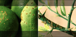
On leaves, first appearance is as oily looking, 2 - 10 mm circular spots, usually
on the abaxial surface (reflecting stomatal entry following rain dispersal).
Lesions are often similarly sized. Later, both epidermal surfaces may become
ruptured by tissue hyperplasia induced by the pathogen.

|

|

|

|

Tell-tale Signs
On leaves, stems, thorns
and fruit, circular lesions become raised and blister-like, growing into white or
yellow spongy pustules. These pustules then darken and thicken into a light tan
to brown corky canker, which is rough to the touch.
|

|
On stems, pustules may
coalesce to split the epidermis along the stem length, and occasionally girdling
of young stems may occur. Older lesions on leaves and fruit tend to have more
elevated margins and are at times surrounded by a yellow chlorotic halo (that
may disappear) and a sunken center. Sunken craters are especially noticeable on
fruits, but the lesions do not penetrate far into the rind. Defoliation and
premature abscission of affected fruit occurs on heavily infected trees.

|

|

|

|

A Closer Look
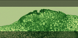
An essential diagnostic symptom is citrus tissue
hyperplasia (excessive mitotic cell divisions), resulting in cankers. In typical
natural infestations, all young aerial parts of the Rutaceous host can be
conspicuously affected.
|

|
Cankers may be
present on leaves, stems and fruit of mature trees; canker symptoms on leaves and
fruit can be readily obtained in artificial inoculations. If cankers are not
present on leaves, stems and fruit of mature trees, or if leaves and fruit of
susceptible citrus species do not develop cankers following artificial inoculation,
a diagnosis of citrus canker is not indicated. This may seem obvious, but a fungal
disease that did not affect fruit was misdiagnosed in Mexico in 1982 as a "form"
of citrus canker, and opportunistic leaf spotting xanthomonads that did not cause
cankers or affect the fruit of mature trees were misdiagnosed in Florida in 1984
as yet another "form" of citrus canker (refer below).

Occurrence of lesions is seasonal, coinciding with periods of heavy rainfall, high
temperatures and growth flushes. These factors generally coincide with early
summer in citrus growing regions where rainfall increases as temperatures increase.
Citrus canker is unlikely to be found in regions where rainfall decreases as
temperatures increase.

Signs of the pathogen should be evident, even in older lesions, as masses of
rod-shaped bacteria streaming from the edges of thinly cut lesion sections. If
bacterial streaming is not observed, a diagnosis of citrus canker is not indicated.

Some confusion is in the literature because the canker-like disease "mancha
foliar", caused by Alternaria limicola [11,16] was found in Mexico in 1982 and
reported to be "citrus bacteriosis" and a "form" of citrus canker [12]. A USDA
embargo on Mexican citrus was the result, despite the fact that Koch's postulates
were inconsistent, lesions were not found on fruit, lesions developed most
strongly in the dry season, and bacterial streaming was not observed [18].
There are no Xanthomonas strains known to cause the symptoms described as
"citrus bacteriosis".

|

|

|

|

Subtle Differences
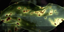
Unlike Citrus Canker, 'nursery leaf spot'
is indicated by flat lesions on the leaf, rather than elevated pustules,
and an absence of fruit or stem lesions.
|

|
Some confusion is in the literature because the opportunistic leaf-spotting
disease "citrus bacterial leaf spot", or "nursery leaf spot" caused by X.
campestris pv. citrumelo [6,7,9] was found in Florida in 1984 and reported
to be a new "form" of citrus canker [15]. A USDA quarantine and grower
compensation program was the result, despite the fact that cankers were
never observed, and mature trees in groves were not affected. Lesions were
found only sporadically in citrus nurseries. By contrast with citrus canker
disease, bacterial streaming is not observed from older lesions of citrus
bacterial spot, even when caused by the most "aggressive" strains of X.
campestris pv. citrumelo.

V. Isolation

VI. Identification

VII. Pathogenicity

VIII. Storage of Organism

IX. Reported Host Range

X. Geographical Range and Spread

XI. Suggested Taxonomic Keys

XII. References

For general inquiries, please send e-mail to
info@ipgenetics.com. For web site errors or content issues, please e-mail
webmaster@ipgenetics.com.

 Copyright © October 2001 Integrated Plant Genetics, Inc. -- All Rights Reserved Copyright © October 2001 Integrated Plant Genetics, Inc. -- All Rights Reserved
|
|

|
|


